Congenital Heart Diseases
Although their frequency has fallen, there are still some birth defects that affect the heart in both children and adults. We are aware of the importance of becoming the allies of parents for curing or providing palliative care for a child with a congenital cardiopathy. This is possible thanks to major advances in treatment.
Congenital heart disease percutaneus treatment
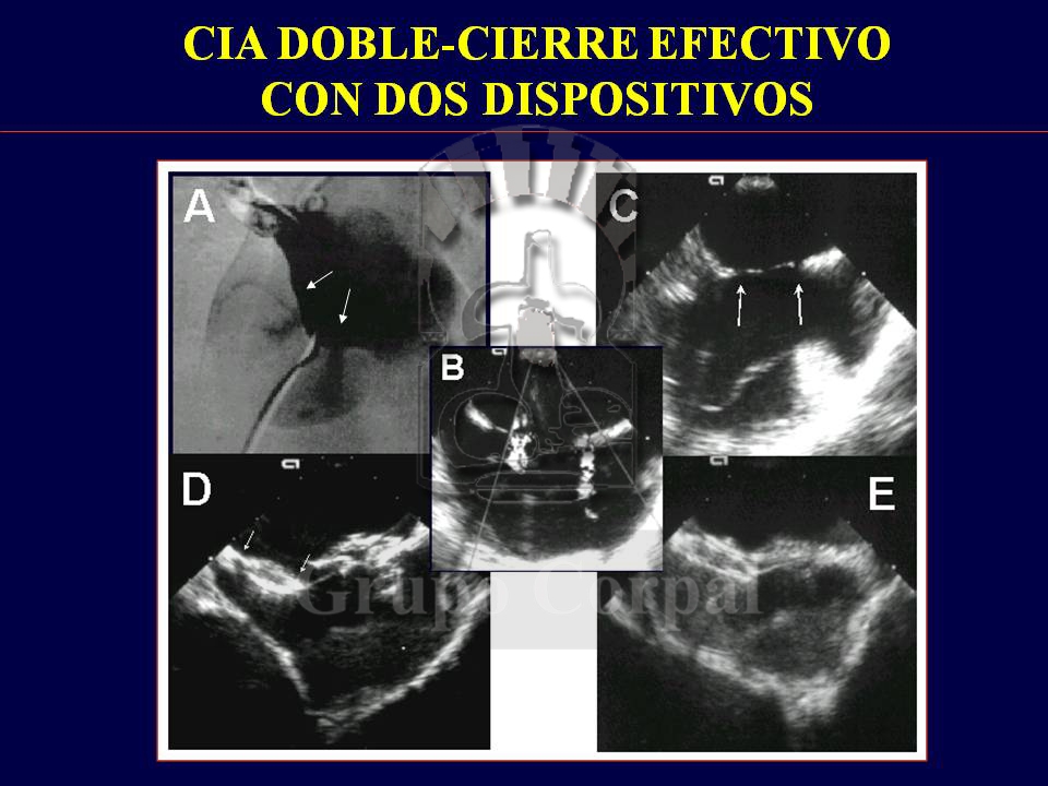
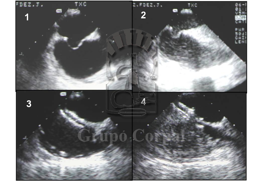
Percutaneous closure of Atrial Septal Defect (ASD)
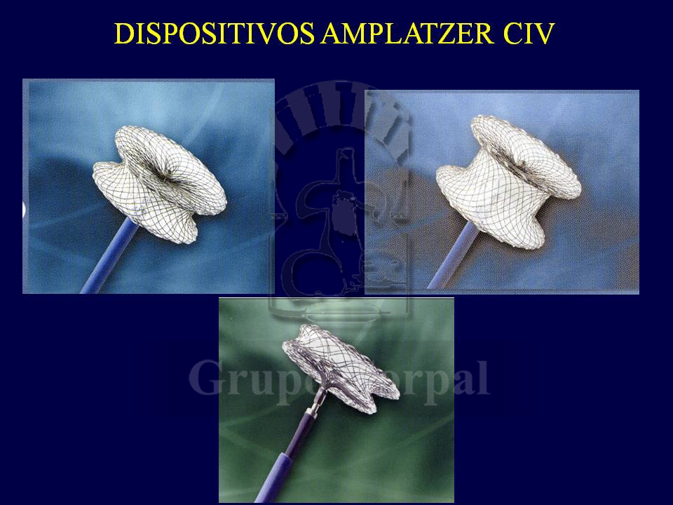
The closure of congenital septal defects has always been a surgical procedure with good results and few complications. Since the 1990s, however, different devices have been designed to be inserted in the circulatory system folded, to occlude and correctly direct the circulation.
Because of the defect, at each heartbeat a jet of blood travels from left to right in the atrium, increasing the blood volume reaching the lungs, causing vascular plethora and, in the long term, heart failure,…
Closure of ventricular septal defect
VSD (ventricular septal defect) is a serious congenital defect that can exceptionally be acquired after rupture of the interventricular septum after acute myocardial infarction.
With this defect, at each heartbeat, a het of blood passes from left to right, increasing the volume circulated to the lungs and leading to vascular plethora and heart failure, as well as the development of pulmonary hypertension.
Patent Foramen Ovale Closure
PFO (patent Foramen Ovale) is a valve mechanism in the interatrial septum; with growth, left atrial pressure usually closes the foramen. However, it is estimated that it remains open during the lifetime of 15% of the population.
The valve mechanism is due to the embryological interposition of the secundum and primum septi, the former growing downwards and the other upwards.
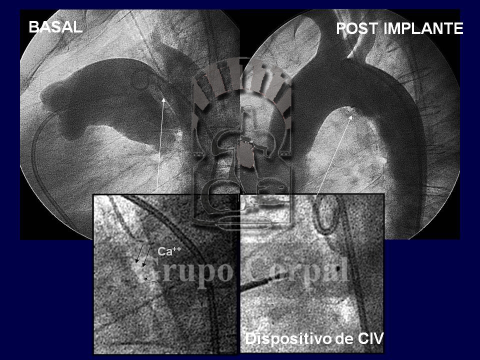
Closure of persistent ductus arteriosus
PDA (Persistent Ductus Arteriosus) is a congenital disorder that, depending on its size, can cause serious problems at any age.
What is normal in intrauterine life and just after birth (conduct connecting both circulations, the aorta and the pulmonary artery), when the lungs expand and circulating oxygen increases, tends to close in the neonatal period due to endo-proliferative stimulation of the conduct together with vasoconstriction by smooth muscle fibres.
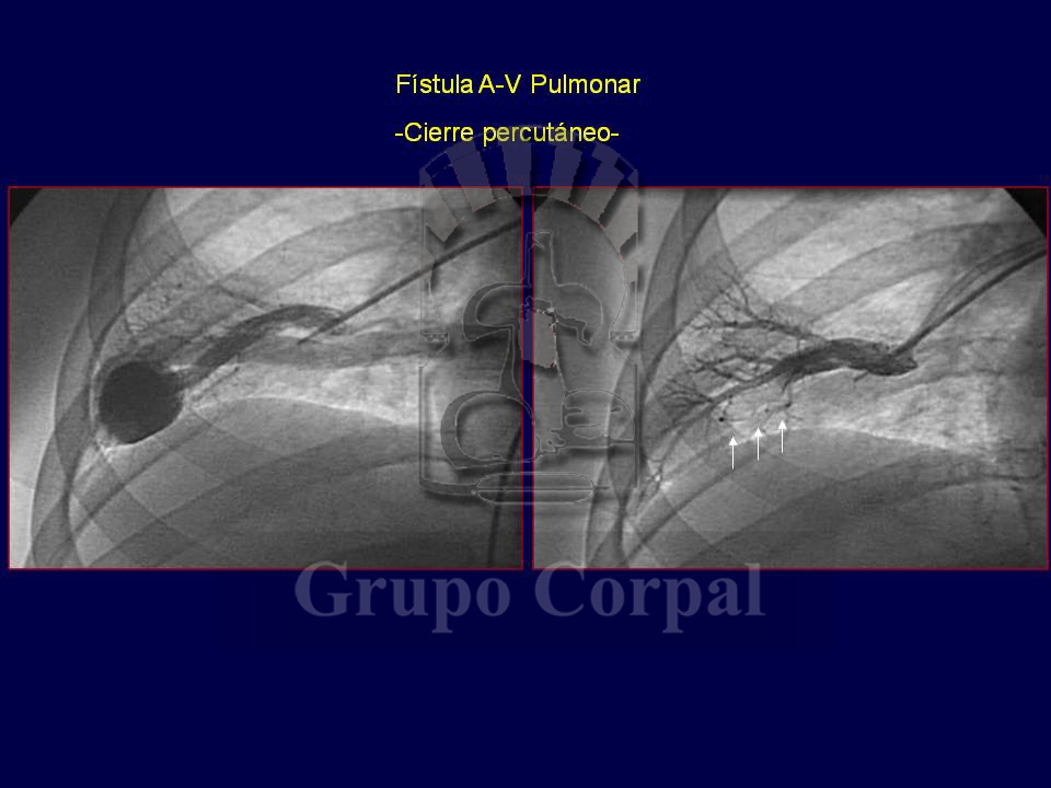
Closure of arterial-venous and venous-systemic fistulas
Fistulas are shunts between arterial and venous systems that prevent capillary circulation and overload venous return.
Arterial-venous fistulas produce hyperkinetic circulatory overload and can be found in any organ or tissue, including the skull of neonates, leading to severe heart failure.
If the fistula is pulmonary (pulmonary artery-vein), it can also enable small clots generated in the venous system to reach the left atrium, and from there to the systemic circulation, with serious repercussions.
Other treatments
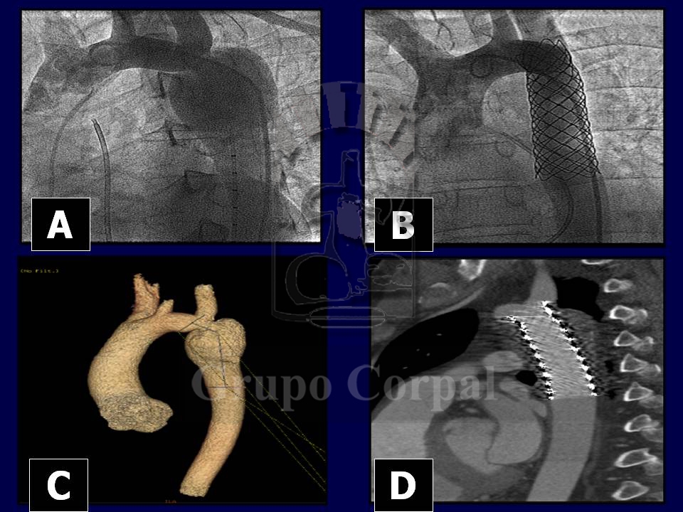
Stent implantation in Coarctation of Aorta
Aortic coarctation is one of the most common congenital cardiopathies. It involves the narrowing of the aortic span at the aortic arch that causes stenosis with cystic necrosis of the middle layer and weakening of the wall.
This systemic stenosis of the most important artery generates hypertension in the top part of the body (left ventricle, head and arms) together with hypoperfusion distal to the coarctation (abdominal organs and legs). The only possible compensation is the development of collateral circulation, which takes time after birth. Its clinical presentation can vary greatly. Read more
Aortic and pulmonary valvuloplasty
Congenital pulmonary and aortic valve stenosis is a serious condition that can have different clinical presentations. Extremely serious cases present in the neonatal period with left heart failure in case of the aortic valve, and right heart failure and hypoxemia in case of the pulmonary valve. Both these conditions are critical and life-threatening without timely mechanical treatment. If the condition is not extreme at birth, compensation mechanisms become gradually established in toddlers’ hearts, guaranteeing blood flow through the stenotic valves.
Right and left ventricular hypertrophy is the main compensation mechanisms for increasing contraction strength. Despite such mechanisms, however, the condition can become more serious over time, leading to intraventricular hypertension, under-development and heart failure. Read more
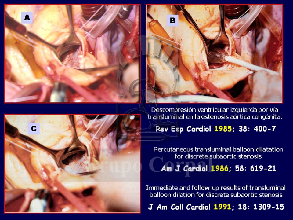
Dilation of Discrete Subaortic Stenosis
Sub-aortic stenosis represents a mechanical obstacle for ventricular ejection located just beneath the aortic valve, in the left ventricle outflow track. It is a rare, mysterious, disease of which we know little.
From an anatomical perspective, on 85% of all occasions the obstructive mechanism is based on a fine sub-aortic membrane that, acting like a diaphragm, produces obstruction to ventricular ejection. The tissue involved is similar to that of the endocardium, and can grow in half-moon shape or create a complete diaphragm. Read more
Percutaneous management of complex congenital cardiopathies
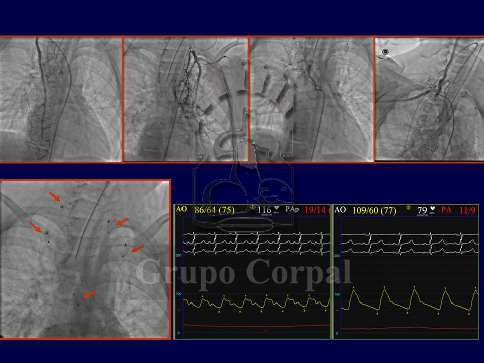
There are complex congenital cardiopathies that require hybrid management of percutaneous procedures to facilitate or complete surgical outcomes. They are medical-surgical programmes that can be fundamental when planning the therapeutic strategy for such cardiopathies in the long term.
The most common are based on cardiopathies involving a “single ventricle” heart that had to be prepared for a Fontan surgery (connecting the vena cavas with the pulmonary artery) Before that indication we have to maintain an appropriate pulmonary flow without exceeding a pressure value that could prevent such a connection. Read more
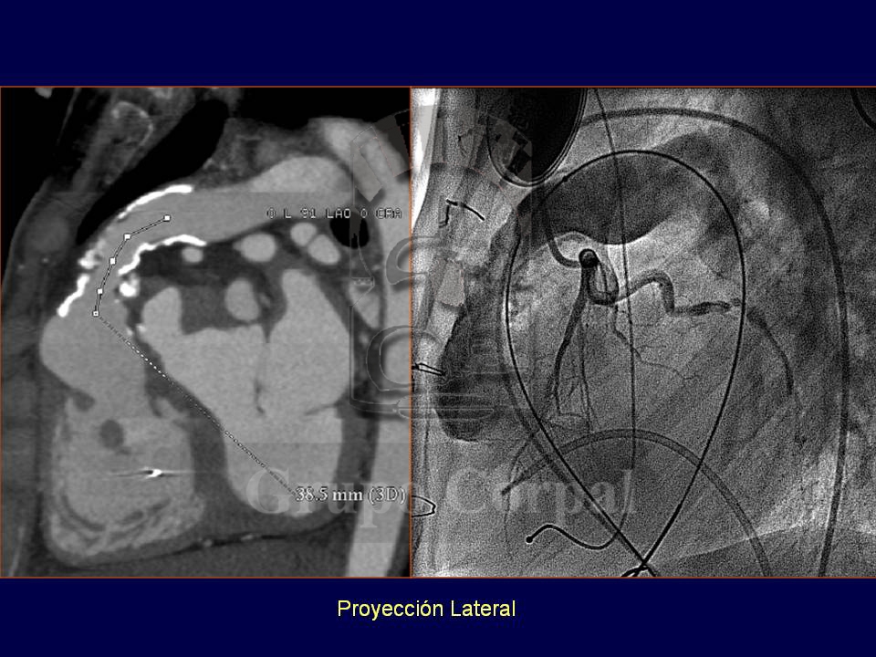
Percutaneous implantation of pulmonary valves
Since the turn of the century it has been possible to percutaneously implant a biological valve in the pulmonary position. What previously required surgery can now be achieved by puncturing the femoral or jugular vein. This transvenous access enables to take a cannula to the pulmonary artery through which a balloon catheter is inserted, installing a stent that, when the balloon is inflated, implants the valve in the correct position.
The first valve was developed by Dr Bonhoffer from the bovine jugular vein, which has leaflets to favour venous return. Read more
